

A rather heated exchange over a new mouse study reporting that an mRNA-vaccine-type lipid nanoparticle increased metastatic burden. Jeffrey Morris called the paper a “potential mechanistic hypothesis.” JikkyLeaks responded that the investigators had actually produced the effect experimentally.
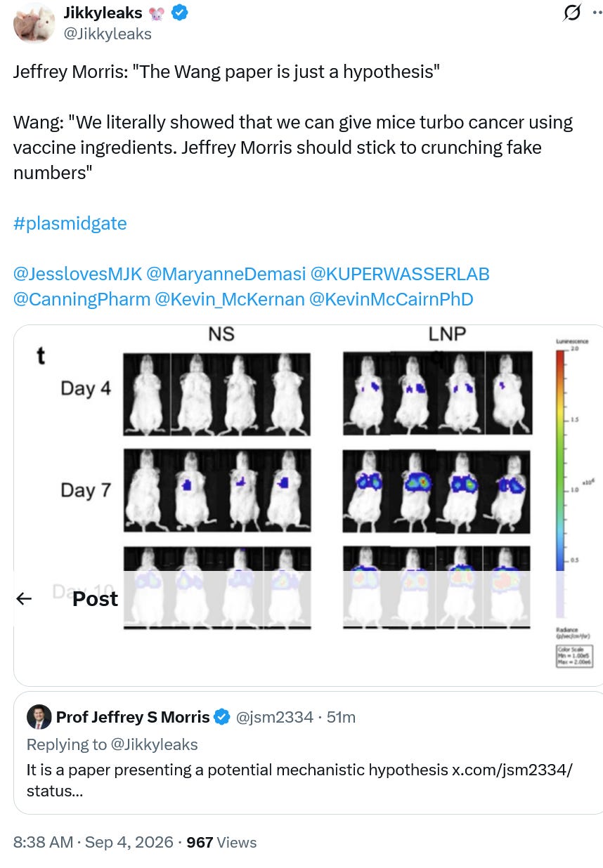
Well, the authors of this paper DID demonstrate increased metastatic burden in several mouse models and performed intervention experiments supporting a biological mechanism. But they did not show that LNPs “gave mice cancer outright.” The mice were deliberately challenged with aggressive tumour cells. Under the experimental conditions, the observed effect was enhanced metastatic seeding, colonization or progression, but not a direct tumour initiation per se. However, this is an important and serious safety signal and was well done with many controls. Importantly, it showed some, but not all, LNP formulations create conditions favourable to metastasis, and this is my biggest takeaway.
Jessica Rose and Geneverter Burger have also weighed in.
The paper is behind a paywall, but I did get access. And because there are a lot of figures but no good graphic abstract, I had SciSpace make me one.
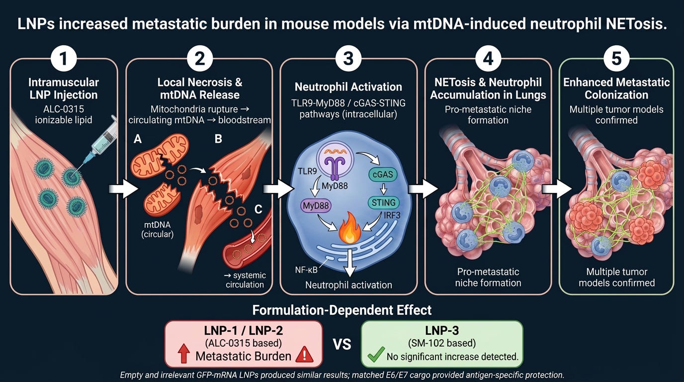
Not its best work, but it is clear.
OK, now to my analysis, such as it is.
What the investigators actually tested
Hybrid Pfizer/Moderna-like LNP-1. The primary formulation contained ALC-0315, the ionizable lipid used in the Pfizer vaccine, together with helper lipid, cholesterol and DMG-PEG 2000, which is the PEG lipid used in Moderna’s formulation rather than Pfizer’s ALC-0159. The paper did not explain this hybrid design.
Furthermore, the lipid molar ratio differed from the published Pfizer ratio. LNP-1 therefore contained more ionizable lipid and helper lipid, less cholesterol, and a different PEG lipid.
If you change the lipid molar ratio, you aren’t just “tweaking a recipe”; it is a fundamental shift in the supramolecular architecture of the particle. You are altering the fluidity, phase behaviour, and electrostatic surface potential of the LNP.
Pfizer-like LNP-2. This formulation contained ALC-0315, helper lipid, cholesterol and Pfizer’s PEG lipid, ALC-0159, at the published Pfizer-BioNTech molar ratio of 46.3:9.4:42.7:1.6.
Moderna-like LNP-3. This formulation contained SM-102, helper lipid, cholesterol and DMG-PEG 2000 at the published Moderna molar ratio of 50:10:38.5:1.5.
LNP-2 and LNP-3 match the commercial products in the lipid components and use the same ratios. But they are not identical because of the manufacturing process and other variables and thus may have different biological “identities.”
The investigators also tested the following mRNA-loaded formulations:
GFP-mRNA-loaded versions of LNP-1, LNP-2 and LNP-3. GFP is an irrelevant reporter protein in these tumour models, making these useful controls for whether loading the LNP with unrelated mRNA altered the result. This is an important control.
An E6/E7 mRNA-loaded version of LNP-1. E6 and E7 are oncoproteins produced by high-risk HPV. The TC-1 mouse tumour cells used in this experiment express HPV16 E6 and E7, allowing the encoded proteins to serve as matched tumour antigens and generate an antigen-specific antitumour response.
The formulation comparison may be the biggest bombshell imho
I am always looking at the supramolecular assemblies and how that contributes to the findings of many studies

Because LNP-1 already contains DMG-PEG 2000, while LNP-2 contains ALC-0159, the shared component is primarily ALC-0315, the ionizable lipid. However, it may not be the ionizable lipid ALONE. Changing an ionizable lipid changes particle formation, RNA association, apparent pKa, membrane interactions, protein corona, intracellular processing, inflammatory activity and biological fate.
There were other formulation issues, including:
different particle sizes, especially after mRNA loading
possible differences in encapsulation and surface presentation;
no matched physicochemical comparison beyond basic size distribution
no head-to-head measurement of muscle necrosis, mtDNA, neutrophils and NETosis for all 3 LNP formulations.
The Moderna-like negative result is meaningful, and the authors did not discuss it. WHY? Genervter Bürger’s Substack has some plausible hypotheses for why we see a difference.
Still, this is a massive “tell.” It proves that the metastatic risk is not an inherent property of “mRNA-LNP technology” as a whole; it is a design and product-specific hazard. The divergence between SM-102 and ALC-0315 suggests that the supramolecular assembly is the true regulatory variable. And no one is measuring it. More on this in this here

and here.

When you change the ionizable lipid (from ALC-0315 to SM-102) and the lipid ratios, you aren’t just swapping a chemical; you are changing the entire soft-matter physics of the particle:
Why did the ALC-0315 formulations increase metastatic burden while the Moderna-like SM-102 formulation did not? The study does not answer this, but several formulation-dependent properties should be examined.
Ionization profile and apparent pKa. ALC-0315- and SM-102-containing particles may have different apparent pKa values and protonation profiles. This could change the number, location and cooperativity of protonated ionizable lipids under extracellular, endosomal and lysosomal conditions.
Membrane fusibility and phase behaviour. The headgroup, linker and hydrophobic tails influence lipid shape, packing, and curvature stress. This means the formulations may differ in membrane insertion, lipid exchange, endosomal escape, lysosomal injury and direct muscle-cell toxicity. This could determine whether an exposure crosses the threshold required for substantial necrosis and mtDNA release.
Biocorona and immune recognition. Following IM administration, the particles enter lymph and plasma. Different surface chemistry and organization could produce different coronas, including differences in complement proteins, immunoglobulins, apolipoproteins and other opsonins. These coronas could alter uptake, clearance, inflammatory signalling, and neutrophil recruitment.
Lipoprotein association and remodelling. LNPs may exchange lipids and apolipoproteins (ApoE or ApoB) or form hybrid lipid assemblies. ALC-0315 and SM-102 formulations could therefore acquire different biological identities, cellular targets and clearance pathways after administration.
Particle size, curvature and structural heterogeneity. The empty SM-102 LNP-3 was substantially larger than the primary ALC-0315 formulation, and loading mRNA altered the sizes of all three formulations differently. Surface curvature, blebs, PEG distribution, and internal lipid–RNA organization may differ. Because dynamic light scattering (DLS) cannot determine these structural properties, the reported particle sizes identify a potential confounder rather than explain the biological result.
RNA association and accessibility. The different size changes after mRNA loading suggest that the formulations reorganized differently around RNA. This is key. Because the empty SM-102 LNP-3 also failed to increase metastasis, RNA encapsulation rate cannot explain the primary formulation-dependent difference.
The authors’ failure to provide a head-to-head comparison of muscle necrosis, mtDNA release, and NETosis across all three formulations is a huge missed opportunity that is hard to explain.
By not doing this, they’ve left us with a important questions and ambiguity.
Is SM-102 “safer” because it causes less muscle necrosis?
Or does it still cause necrosis, but the resulting “debris” doesn’t trigger the TLR9/cGAS-STING pathway?
Or does it cause necrosis outside the time windows of the study?
We now have good data that the SM-102/Moderna-like architecture behaved differently in this metastatic model than the Pfizer-like architecture. If this is true, we should expect to see various studies and commentaries on exactly why SM-102 didn’t trigger the metastatic cascade. We shall see.
The DiD biodistribution experiment: following lipid-associated signal rather than cargo
The investigators did something interesting with the biodistribution study. Most LNP biodistribution experiments follow only one component, such as the mRNA, luciferase expression as a proxy for protein expression, or one particular lipid. The results are usually discussed as though the entire intact particle had been followed. It has not.
Here, the authors incorporated DiD, a highly lipophilic fluorescent dye, into the LNP lipid phase and compared DiD@LNP with free DiD. Because DiD partitions into the lipids, it follows DiD-associated lipid material rather than the mRNA cargo. However, it does not identify which of the four LNP lipids it accompanies, nor does it indicate whether the original intact LNP remains around it.
The whole-animal DiD@LNP signal appeared to become progressively stronger in the abdominal region through 48 hours. At 48 hours, ex vivo organ imaging showed that the signal was predominantly hepatic and significantly greater in the liver than after free DiD administration. Lung fluorescence did not differ significantly between free DiD and DiD@LNP.


The lack of elevated DiD signal in the lung is consistent with the authors’ proposed mechanism. They proposed that large quantities of intact LNP DID NOT accumulate in the lung. They proposed a relay: local muscle injury released a circulating damage signal (mitochondrial DNA or mtDNA), which then produced distal lung inflammation. The LNP was administered intramuscularly, matching how the vaccine is given.
I would add another possibility. Circulating mtDNA doesn’t need to be free mtDNA. Some could be carried in EVs, including exosomes, along with other cellular components and possibly LNP-derived material. The investigators did not separate free from protein-bound or extracellular-vesicle-associated mtDNA, so this remains unanswered. I discuss this more later.
The mechanistic chain
See my graphical abstract at the start of this post for an overall view.
This is a dense paper, so I used ChatGPT to help organize the experimental findings into this stepwise sequence, which I then checked against the paper.
OK, so if you think about it step by step, this means:
Intramuscular LNP injection caused local muscle injury and necrosis.

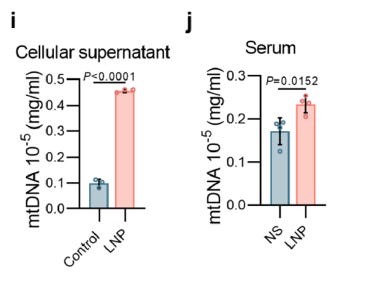
Damaged muscle cells released mitochondrial material, including mtDNA
Serum mtDNA increased after injection.
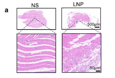
Circulating inflammatory signals recruited neutrophils and cells identified as myeloid-derived suppressor cells. These closely related, sometimes overlapping myeloid populations can suppress anti-tumour immunity and help establish a tumour-permissive environment.
TLR9–MyD88 and cGAS–STING signalling participated in the response.
Neutrophils accumulated in the lung and acquired markers consistent with an N2-like, pro-metastatic phenotype.
NETosis released DNA, myeloperoxidase, neutrophil elastase and histones.
Positively charged histones increased adhesion between tumour and endothelial cells, apparently through interactions with negatively charged sialic acid and heparan sulfate.
The authors propose that this increased tumour-cell adhesion facilitates metastatic capture, retention or colonization in the lung.
This is a specific mechanistic sequence and claim rather than the general “inflammation promotes cancer.” What I find especially interesting is the final step regarding adhesion between tumour and endothelial cells. Histone H3 appears to act as an electrostatic bridge. Spike protein might contribute to such interactions, as might a protonated ionizable lipid. The present study did not examine either possibility, so these are just questions I have.
Also, most experiments supporting the mechanistic claim use LNP-1, the Pfizer-like LNP with Moderna’s PEG and different molar ratios or percentages of the component lipids. Thus, these results may not track exactly with the commercial products, though there is enough here to insist on the appropriate studies. Would the results be different otherwise? It would have been very helpful indeed, if the Moderna-like LNPs were also studied in the same experiments. Sigh.
Why the controls are unusually good
One strength of the Wang et al. paper is its rigorous control experiments. This makes it quite dense to read. They systematically eliminated "confounders" (alternative explanations) that industry defenders usually use to dismiss such findings. These included:
Untreated and saline-injected controls distinguished LNP effects from needle injury. If the metastasis were just from the physical act of injecting a needle, the NS group would have shown the same effect as the LNP group. They didn't. This proves the effect is chemically mediated by the LNP. (someone on X tried to float that needle injury canard!)
To rule out "Payload-induced" effects: They compared Empty LNPs (eLNPs) vs. GFP-mRNA-loaded LNPs. Both caused the same metastatic promotion. This is the "smoking gun" that proves the LNP delivery vehicle itself is the bioactive driver, not the mRNA or the resulting protein. This matters because some e-LNP studies show that mRNA itself is an additional source of inflammatory signal (maybe due to contaminating dsRNA, endotoxin or dsDNA).
To prove "Antigen-Specific" efficacy vs. "LNP-induced" danger: They used an E6/E7 mRNA vaccine arm (for HPV-related tumors). This arm showed protection against tumours. This shows the LNP is a “double-edged sword” as we have been saying for a long time. There is no doubt mRNA vaccines elicit an immune response, but this is at the cost of systemic inflammatory/pro-metastatic damage. The platform carries an inherent toxicological “tax.” Thus, the push to make “safer” LNPs.
They performed a “Robustness Assessment.”
LNPs given before and after tumour-cell inoculation tested two different phases. This proves the LNP effect isn’t just about “priming” the body before the cancer arrives; it is capable of accelerating existing metastatic disease. This is especially relevant for patients who might already have subclinical or established tumours. And this is what clinicians see.
A clear dose-response relationship was seen, supporting biological plausibility, a critical finding because the doses used were large, which many critics have pointed out. Nonetheless, the fact that the metastatic effect scaled with the lipid mass is the hallmark of a true biological effect rather than an experimental artifact. This follows classic toxicology principles to show a causal link. I talk about this in more detail below.
They used three distinct mouse tumour models: B16-F10, 4T1 and CT26, reducing reliance on a single tumour line. Showing the same pro-metastatic effect across such different tumour types suggests a platform-wide risk and is model-agnostic, not a quirk of a specific cancer.
They didn’t look at just one organ, like the lung. The effect was observed in spontaneous breast-cancer metastasis and liver-directed metastasis. The liver has a completely different environment than the lung, so this implies systemic “pro-metastatic” change.
To confirm the "Biological Pathway,” they used Knockout Mice (Tlr9-/-, MyD88-/-, Sting-/-). They showed that removing these specific inflammatory sensors abrogated the effect. It is not just general "inflammation" but specifically mtDNA-triggered TLR9/cGAS-STING signaling. They also looked at Gene Ontology enrichment of signalling pathways and found genes such as Cxcl2, S100a8, and Cxcr2 highly expressed, which are potent chemotactic mediators for neutrophils.
This is particularly interesting. See Genervter Bürger Substack for more, especially on S100a8. They also showed the following:
Neutrophil depletion reduced metastatic burden, confirming neutrophils’ role.
Cl-amidine and DNase I interventions confirmed and supported a role for NETosis.
Exogenous mtDNA partially reproduced the phenotype.
The histone/endothelial-cell experiments supplied a plausible final adhesion mechanism.
A Pfizer-like and Moderna-like formulation comparison revealed formulation dependence. This is perhaps the most bombshell result, imho, as I discussed earlier. It suggests safety is not a property of the "LNP" label as an inert truck. It points to specific lipid chemistry and the supramolecular assembly. The fact that one formulation triggered this pathology while another did not is a massive regulatory red flag.
No single experiment proves the full chain, but the authors did far more than observe additional lung nodules and then attach a speculative explanation afterward.
7. What the study did not establish
It did not show cancer initiation
The mice received tumour cells deliberately. The outcome was metastatic burden, not transformation of normal cells or spontaneous tumour formation. Still, metastatic burden is not a small thing. At all.
The challenge models are aggressive
As I understand it, tail-vein and intrasplenic tumour-cell injection bypass much of the natural metastatic cascade (just like vaccines do, lol). They are useful for studying survival, adhesion and colonization of circulating tumour cells, but they do not recreate the full development of human cancer. So we need to do more studies.
The dose needs context
OK, the pharmacist geek is coming out. The highest reported lipid dose was 12 mg/kg, equivalent to an LNP quantity carrying approximately 10 µg mRNA per mouse. Critics say this is unphysiological and “too high.” However, the study used doses from 1.2mg/kg to 12mg/kg and you can see effects even at the lowest dose.
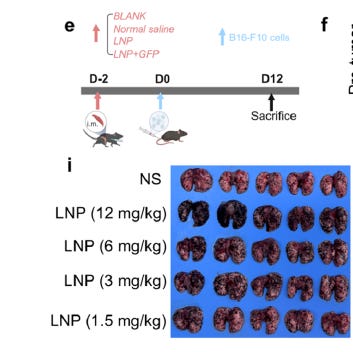
But even with allometric scaling, which regulators use for safety, these findings don’t disappear. If you use the standard mouse-to-human conversion factor of 12.3 (based on Body Surface Area), a 1.2 mg/kg mouse dose scales to roughly 15 mg/kg in a human. But this assumes the LNPs distribute evenly throughout the body. The LNPs do not. This is a mistake many people make when discussing the pharmacological and toxicological effects of these products.
LNPs are designed to target tissues and avoid general systemic distribution. So LOCAL concentration is what matters. The toxicity observed in this study (muscle necrosis, mtDNA release) is concentration-dependent, not body-weight-dependent. Whether it’s a mouse leg or a human arm, the lipid mass is being injected into a very small, confined volume of muscle tissue.
Plus, as Genervter Bürger and I have often discussed, our cell membranes detect particle count. And particle count does not scale allometrically. If you inject a high concentration of nanoparticles into a small muscle, you will cause local necrosis, whether that muscle belongs to a 20g mouse or a 70kg human.
We need a proper dose bridge that accounts for the local effects of LNP particles. Total-body-weight math isn’t going to do it. Furthermore, a single-dose comparison is not sufficient. Each dose triggers a window of inflammation and NETosis and a possible metastatic window. Finally, we need to account for lipid persistence, which we discuss in our L-DMD paper.
The mechanistic pathway is strong, but I have questions
The paper connects muscle necrosis, serum mtDNA, innate signalling, neutrophils and NETosis. But it does not fully establish:
whether circulating mtDNA was free, protein-bound or extracellular-vesicle-associated.
whether mtDNA itself was the principal circulating mediator or if other mitochondrial DAMPs contributed;
whether lipid-associated material, cytokines or EVs also participated;
whether preventing muscle necrosis upstream would abolish the distal effect (does Onpattro, given IV, cause this pro-metastatic state? This would be a very interesting result if not);
why the ALC-0315 formulations differed from the SM-102 formulation.
DNase I also acts on NET DNA downstream, so its protective effect does not by itself prove that circulating mtDNA was the indispensable upstream mediator. It could be that mtDNA is just a marker, and the real “driver” is the LNP-lipid-protein-EV/exosomes, which also trigger mtDNA release.
Our L-DMD paper explicitly discusses how LNPs remodel into hybrid lipoprotein particles and how EVs carry LNP-derived lipids. If the mtDNA is hitchhiking inside an EV along with LNP-derived lipids, then the LNP isn't just acting on the muscle to cause necrosis, it is the carrier of the secondary signal. And after my Exosomes, Schmectasomes Substack post, I am more convinced that unless EVs/exosomes are taken into account, these results may not show the full mechanisms.

Of interest, Wang et al. showed a relatively modest LDH response, which is particularly relevant here because it argues against equating extracellular mtDNA with widespread lytic cell death. This supports the hypothesis that mtDNA may be released through non-lytic or regulated mechanisms associated with cellular stress, membrane dysfunction or altered vesicular trafficking. Please see Falko’s Substack on this particular point.
9. The largest unanswered question: duration
One thing that really stuck out at me was the mismatched time clocks or windows:
The Inflammatory Clock: Neutrophil and MDSC activation is measured in hours (peaking at 12–24h, stopping at 48h).
The Material-Fate Clock: Lipid-associated fluorescence (DiD) is tracked only through 48 hours.
The Metastatic Clock: Tumour burden is assessed weeks later (12–28 days).
This disconnect creates a gap. The study shows an acute window of enhanced susceptibility, but it doesn’t tell us when that window closes. Does the risk vanish at 72 hours? Or are we creating a chronic state of susceptibility? Given the potential risk of lipid persistence, we need to understand this.
A proper follow-up would challenge the mice with tumour cells at 2, 7, 14, and 28 days post-LNP injection. But even more important is the repeat-dose experiment. We are no longer vaccinating in a pandemic. The model is now moving toward a multi-dose, seasonal paradigm. If you trigger a pro-metastatic inflammatory window every few months, are you potentially maintaining a permanently primed pro-metastatic milieu?
10. Summary
This paper does not establish that an authorized mRNA vaccine CAUSES metastatic cancer in people. But this whole causality issue is a trap. Proving causality is almost impossible. But you can define if it is attributable and how strong that attributability is. This paper does establish that an LNP assembly can be biologically active independently of its encoded cargo (as we have been saying for ages), can injure tissue at the injection site, can initiate systemic signalling, and can alter a distant tissue environment in a way that favours metastatic colonization.
Just as importantly, a change from an ALC-0315 formulation to an SM-102 formulation changed the result. Safety therefore does not belong to the generic label “LNP.” It belongs to the precise molecule, its supramolecular assembly, its dose, its route, its biological transformations and its complete fate.
The message is not “vaccine ingredients give mice turbo cancer” or “it is just a hypothesis.” The paper presents substantive preclinical evidence of a formulation-dependent biological effect, and more than enough reason to ask the next set of experiments.
If a substantive signal like this emerged during conventional drug development, especially for a product intended for patients with cancer, it would ordinarily demand some kind of resolution or explanation before clinical advancement, not just more papers.
To be specific: if a candidate drug’s preclinical package showed dose-dependent enhancement of metastatic burden across several tumour models, accompanied by a plausible mechanistic chain and partial reversal through genetic or pharmacological interventions, well, one would expect a very serious development hold by the manufacturers themselves, let alone the regulators.
Especially here, because:
the effect appeared with both empty and GRP or irrelevant-mRNA-loaded particles;
it occurred when LNP was administered before or after tumour-cell inoculation;
it appeared in multiple tumour models and at more than one metastatic site;
the formulation caused local necrosis and distal lung inflammation;
neutrophil depletion and NET-directed interventions reduced the effect;
one composition produced the effect while another apparently did not.
MY CONCLUSIONS
These data do not establish the magnitude or specific risk of LNPs in humans, but they are sufficient to make regulatory inaction untenable, imho.
Formulation-specific studies examining tissue injury, biological fate, mtDNA release, myeloid activation, NETosis and tumour progression should now be required before any new mRNA vaccine, cancer vaccine or other LNP-based therapeutic proceeds clinically. Plus all the other stuff that should have been done in the very beginning for these LNPs (cellular processing, true biodistribution, secondary pharmacology, etc etc)
Existing vaccine LNP formulations should be urgently reassessed against the same questions that we and others have raised for the past four years, with the evidence and regulatory reasoning made public. Here is our submission to ACIP in Sept 2025.
In light of the totality of unresolved evidence, not just this study, regulators should also formally consider whether continued unrestricted use, targeted restrictions, or a temporary hold is warranted while these gaps are addressed.
Enough.
Pray the rosary, things are getting heated
Thanks for reading.
We need to know what happens when LNP are converted into EV/HDL/LDL and their bio distribution? Can they then pass the BBB? What scout genotoxic testing? There is zero animal or human testing to see if mRNA can get into the nucleus? We need more than verbal assurances, we need DB testing, if there is any danger of passing this to future generations? And what about ribosomal frame shifting from pseudo uridine? What aberrant proteins are produced and can they cross the BBB? What about antibody dependent enhancement and immune imprinting? Did the mRNA vaccines be cause mutations like omeron from selective pressure? Did 1.1 million Americans die by releasing a vaccine in the middle of a raging pandemic? Vaccines do cure anything, they are designed to prevent disease, not cure it!
Danke